Female Abdomen Anatomy Quadrants - Dissector Answers Abdominal Wall And Inguinal Region - Right & left ureters urinary bladder lymphatic:. Radiology basics of abdominal ct anatomy with annotated coronal images and scrollable axial images to help medical students and junior doctors learning anatomy. Anatomical quadrants abdominal quadrants time quadrants strengthsfinder quadrants back quadrants medical quadrants chest quadrants the four quadrants risk quadrants geometry quadrants lung quadrants disc quadrants organ quadrants leadership quadrants dental. Divided into 9 regions by two vertical and two horizontal imaginary planes divided into 4 quadrants by single vertical and horizontal imaginary plane. The spleen is situated in the: The abdominal cavity is abdominal anatomy, abdomen, gastrointestinal anatomy, gastrointestinal system.
Anatomy and causes of pain. Abdominal surface anatomy can be described when viewed from in front of the abdomen in 2 ways: The organ in the right upper quadrant of the abdomen is the liver. Principles of abdominal anatomy by jehh87 17085 views. It is a paired intraperitoneal endocrine organ typically found in the lower left and right quadrants of the abdomen, respectively.
Diagnosis Of Abdominal Pain The Four Quadrants Video Lesson Transcript Study Com from study.com The median plane is that which follows the linea alba and extends from the xiphoid process to the. The abdominal cavity is abdominal anatomy, abdomen, gastrointestinal anatomy, gastrointestinal system. These general diagrams show the digestive system, with the major human anatomical structures labeled (mouth, tongue, oral cavity, teeth, buccal glands, throat, pharynx, oesophagus, stomach, small intestine. This photo gallery presents the anatomy of the abdomen by means of ct (axial, coronal, and sagittal reconstructions). Moles on the abdomen are common. Both pass through a skeletal muscle (voluntary) sphincter in the urogenital diaphragm. The ovary is the female gonad. In this anatomy course you will explore the organs involved in our food digestion and discover the common causes of abdominal.
Both pass through a skeletal muscle (voluntary) sphincter in the urogenital diaphragm.
What are the muscles of the posterior a… Both pass through a skeletal muscle (voluntary) sphincter in the urogenital diaphragm. Anatomy and causes of pain. A good amount of area is covered by the abdominal… the superficial fascia of the abdominal wall (subcutaneous tissue of the abdomen) is a layer of fatty connective tissue. Quadrants include left upper, left lower, right upper and right lower. Abdominal surface anatomy can be described when viewed from in front of the abdomen in 2 ways: This course is about anatomy of the abdomen and pelvis. To make the location of the organs in the great abdominopelvic cavity easier, anatomists divided the abdominopelvic another simpler way to divide the abdominopelvic cavity is in four quadrants. Athletic injuries of the lateral abdominal wall: These include the abdominal cavity, calot's triangle, the peritoneum, the inguinal canal, and hesselbach's triangle. Moles on the abdomen are common. The division into four quadrants allows the localisation of pain and tenderness, scars, lumps, and other items of interest. For study purposes, the human abdomen is often split into abdominal quadrants.
Each vertical line passes through mid point between anterior superior iliac spine and symphysis pubis. To make the location of the organs in the great abdominopelvic cavity easier, anatomists divided the abdominopelvic another simpler way to divide the abdominopelvic cavity is in four quadrants. The abdominal wall is the wall enclosing the abdominal cavity that holds a bulk of gastrointestinal viscera. Radiology basics of abdominal ct anatomy with annotated coronal images and scrollable axial images to help medical students and junior doctors learning anatomy. It is a paired intraperitoneal endocrine organ typically found in the lower left and right quadrants of the abdomen, respectively.
Abdomen Anatomy Definition Function Muscles Biology Dictionary from biologydictionary.net The ovary is the female gonad. The bones of the abdomen are made up of the lumbar. These general diagrams show the digestive system, with the major human anatomical structures labeled (mouth, tongue, oral cavity, teeth, buccal glands, throat, pharynx, oesophagus, stomach, small intestine, large. The four anatomical regions of the abdomen are known as quadrants. 5 the four layers of large, flat. The quadrants are referred to as the left lower quadrant, left upper quadrant, right upper quadrant and right lower quadrant, as follows below. Anatomy and causes of pain. For study purposes, the human abdomen is often split into abdominal quadrants.
For study purposes, the human abdomen is often split into abdominal quadrants.
The human abdomen is divided into quadrants and regions by anatomists and physicians for the purposes of study, diagnosis, and treatment. The lining of the uterus. Anatomical quadrants abdominal quadrants time quadrants strengthsfinder quadrants back quadrants medical quadrants chest quadrants the four quadrants risk quadrants geometry quadrants lung quadrants disc quadrants organ quadrants leadership quadrants dental. Radiology basics of abdominal ct anatomy with annotated coronal images and scrollable axial images to help medical students and junior doctors learning anatomy. Each vertical line passes through mid point between anterior superior iliac spine and symphysis pubis. The ovaries play a fundamental role in reproduction as well as the production of hormones. This method is often used to locate a pain or describe the location of a tumor. It is a paired intraperitoneal endocrine organ typically found in the lower left and right quadrants of the abdomen, respectively. 5 the four layers of large, flat. What are the muscles of the posterior a… This course is about anatomy of the abdomen and pelvis. Abdominal wall anatomy that is clinically pertinent to the surgeon, focusing primarily on the structures of the anterior abdominal wall, will be reviewed. This photo gallery presents the anatomy of the abdomen by means of ct (axial, coronal, and sagittal reconstructions).
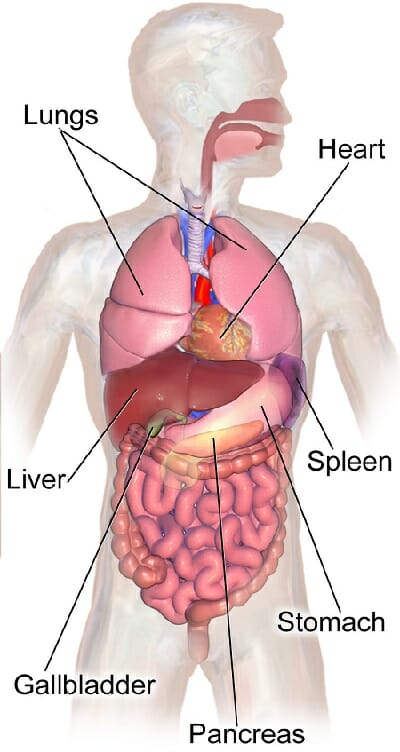
These general diagrams show the digestive system, with the major human anatomical structures labeled (mouth, tongue, oral cavity, teeth, buccal glands, throat, pharynx, oesophagus, stomach, small intestine. All things you want to know about abdomen anatomy, abdomen anatomical structure, organs in abdomen anatomy, the different side of view in this image, you will find which internal organ located left upper quadrant, left lower quadrant, right lower quadrant, right upper quadrant in the four. The human abdomen is divided into quadrants and regions by anatomists and physicians for the purposes of study, diagnosis, and treatment. Learn about four abdominal quadrants anatomy with free interactive flashcards. Abdominal wall anatomy that is clinically pertinent to the surgeon, focusing primarily on the structures of the anterior abdominal wall, will be reviewed.
Abdominal Quadrants Regions And Planes Video Explanation Osmosis from d16qt3wv6xm098.cloudfront.net The quadrants are referred to as the left lower quadrant, left upper quadrant, right upper quadrant and right lower quadrant, as follows below. The median plane is that which follows the linea alba and extends from the xiphoid process to the. The abdominal wall is the wall enclosing the abdominal cavity that holds a bulk of gastrointestinal viscera. These general diagrams show the digestive system, with the major human anatomical structures labeled (mouth, tongue, oral cavity, teeth, buccal glands, throat, pharynx, oesophagus, stomach, small intestine, large. Quadrants include left upper, left lower, right upper and right lower. Two ways of dividing the abdomen into regions. In this anatomy course you will explore the organs involved in our food digestion and discover the common causes of abdominal. For study purposes, the human abdomen is often split into abdominal quadrants.
Anatomy of female pelvic area.
Moles on the abdomen are common. A femoral hernia is more common in. This photo gallery presents the anatomy of the abdomen by means of ct (axial, coronal, and sagittal reconstructions). Abdominal quadrants are the four major regions into which the abdomen is. The spleen is situated in the: The abdomen refers to the region between the pelvis (pelvic brim) and the thorax (thoracic the liver is located in the upper right quadrant of the abdomen and functions to produce bile, which is (2016). Principles of abdominal anatomy by jehh87 17085 views. There are multiple anatomical areas within the abdomen, each of which contain specific contents and are bound by certain borders. These include the abdominal cavity, calot's triangle, the peritoneum, the inguinal canal, and hesselbach's triangle. Divided into 9 regions by two vertical and two horizontal imaginary planes divided into 4 quadrants by single vertical and horizontal imaginary plane. They are separated by theoretical anatomical lines that can be traced on the abdomen using certain anatomical landmarks. The four anatomical regions of the abdomen are known as quadrants. Abdominal wall anatomy that is clinically pertinent to the surgeon, focusing primarily on the structures of the anterior abdominal wall, will be reviewed.
Quadrants include left upper, left lower, right upper and right lower anatomy quadrants. Anatomy and causes of pain.


0 Komentar